Sunil Mamtora1, Oliver Riley1, Jordan Chervenkoff1, Asif Kiani1, Alexander Chiu2, Jonathan Boulton1, Jonathan Luck1 1. Royal United Hospital, Bath 2. Royal College of Ophthalmologists
Introduction- Hospital eye casualty services have remained open throughout the pandemic. There has been a drive to safely minimize face-to-face consultations where possible to reduce the number of hospital attendances.
- The majority of presentations to emergency eye services has been shown to be secondary to anterior segment pathology. We evaluated a smartphone lens attachment (Quickvue, Visuscience Ltd., Shanghai, China) as an adjunct to triage in our eye casualty service.
| 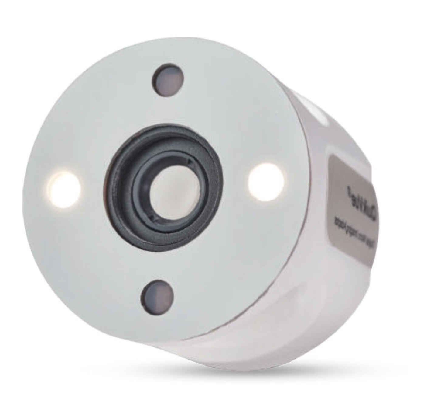 | Figure 1: Picture of lens attachment as used in our study |
|
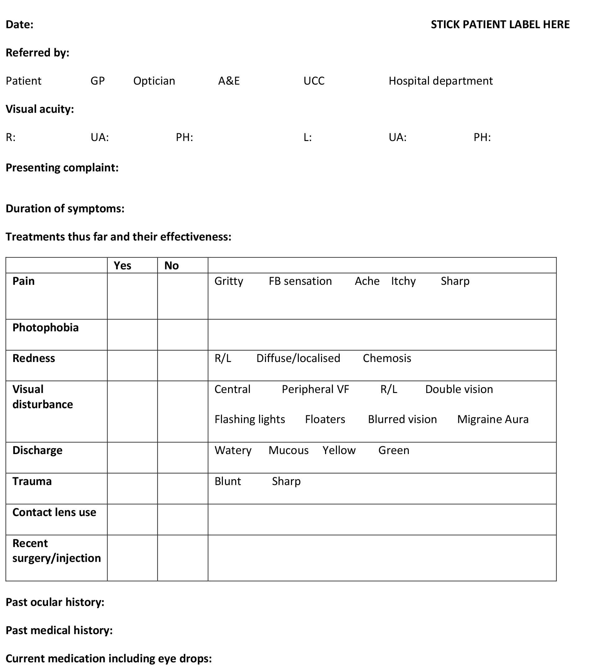  | Figure 2: Questionnaire as used in our study |
| Methods - Anterior segment photography with white and blue illumination was captured and a questionnaire simulating the level of information that could be expected in a referral was completed.
- Patient diagnosis, the appropriate timeframe within which the reviewing clinician felt that the patient could safely have been reviewed within and whether the patient could have been managed without attending the hospital were also recorded.
|
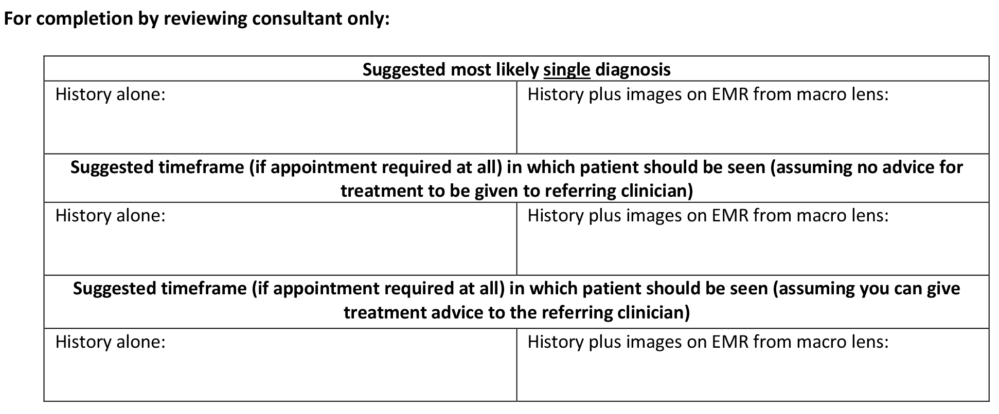
Results - An independent Consultant Ophthalmologist subsequently reviewed the questionnaire only, documenting the most likely diagnosis, the time-frame in which they would review the patient and their confidence in the decision. They then repeated this process with the addition of photographs.
- Diagnostic accuracy was 98.15% with photographs and questionnaires compared to 48.15% with questionnaires alone. The time-frame within which patients would have been reviewed in was significantly longer with photographs and questionnaires compared to questionnaires alone (p<0.05). Clinical confidence in the decision making process was significantly greater with the addition of photographs (p><0.05).
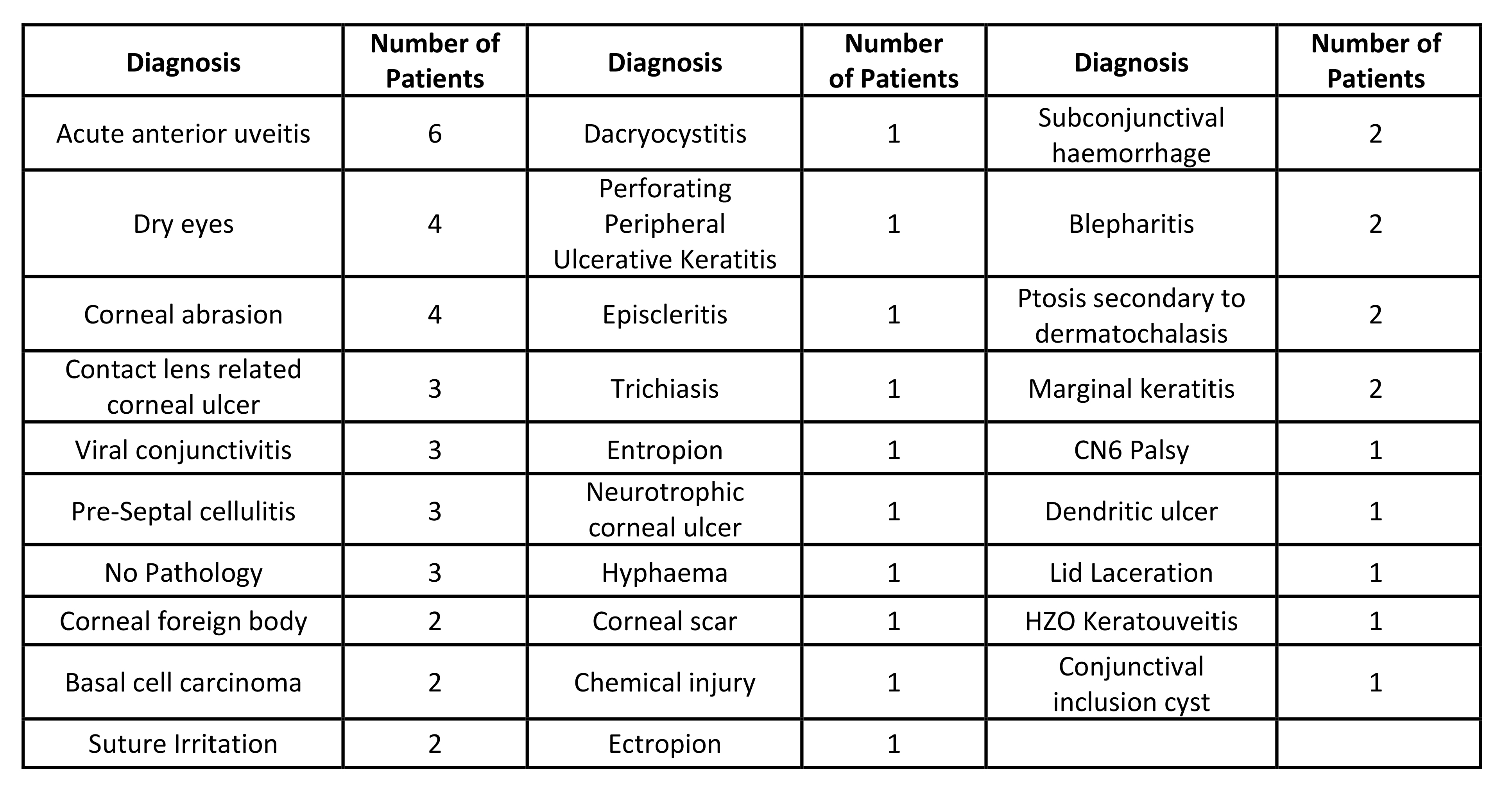
| Figure 3: Diagnoses of the 54 patients included in our study |
|
Discussion - Smartphone based anterior segment photography was shown to have remarkable diagnostic accuracy. This was helped with the presence of both blue and white light photographs for each patient.
- Image capture was made directly to the patient’s electronic medical record in our study using the PowerChart Capture iOS application.
- Potential sources of bias include the knowledge of the photographer of the clinical diagnosis which might have led them to accentuate the relevant clinical findings in the photographs they captured.
- Minimal end user training is required to use this device. Patient positioning requirements are eliminated and this device is particularly useful for ward reviews or in bed-bound patients.
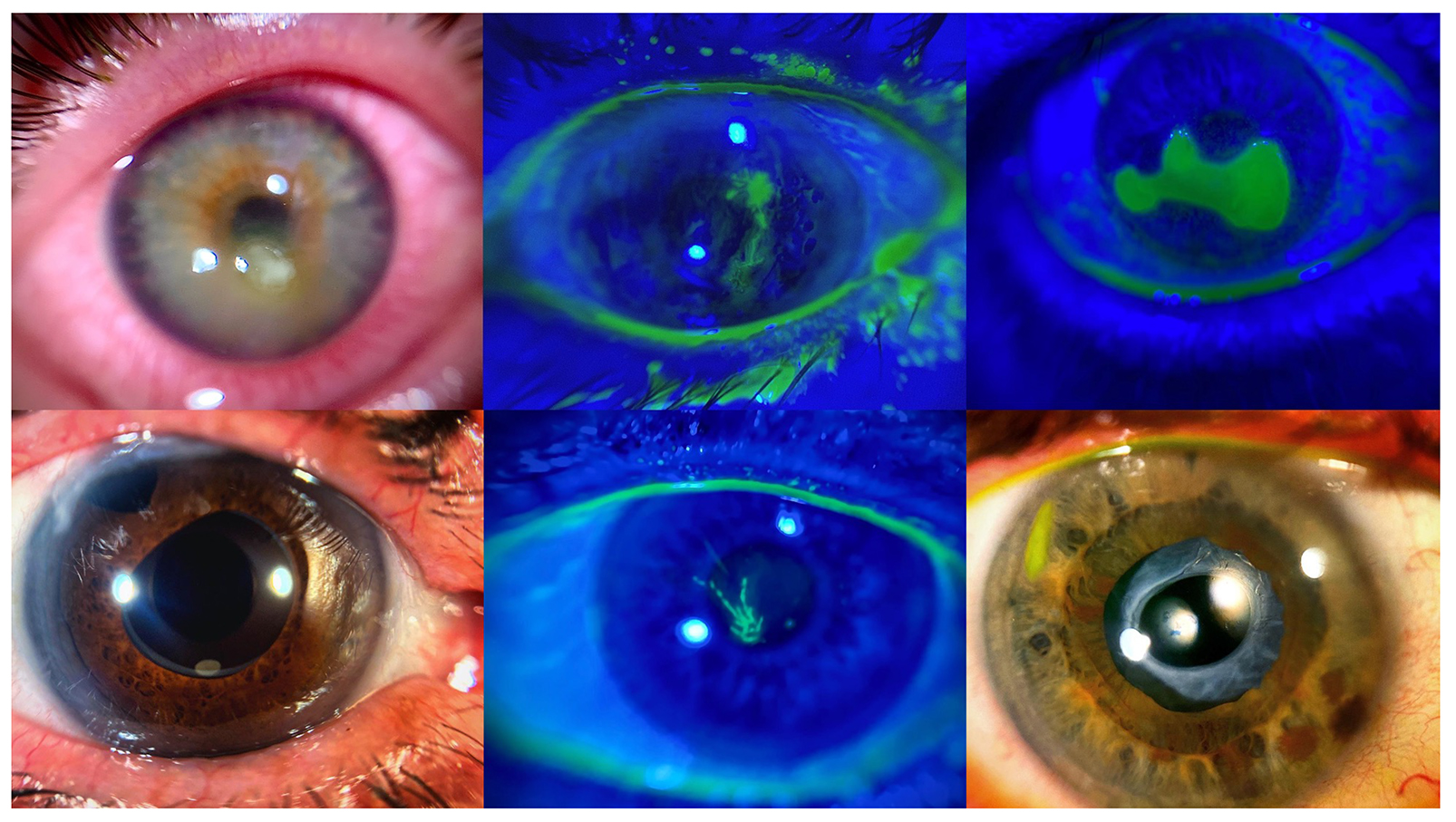
| Figure 4: Sample images as captured during our study |
Conclusions - Smartphone anterior segment photography is an effective adjunct to conventional triage. Beyond the pandemic, it may prove useful in managing the ever increasing demands faced by emergency eye services.
- Coupling this technology with handheld non-mydriatic fundus photography, whose image quality has vastly improved in recent years, provides non-ophthalmologists with a toolkit to perform reasonably comprehensive, digital eye examinations.
Click here to view PDF file. |
|
