




QuikVue Vet Case Share - corneal edema
We are glad to share a vet case study captured by QuikVue eye imaging adaptor from Dr. Allison Fuchs.
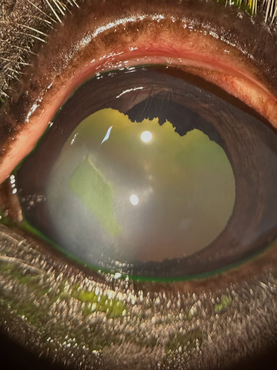
This is a tough case! This is a horse with corneal ulcers that keep coming back over an area of corneal edema, which just means fluid in the cornea. They have been battling this for several years and we just started seeing him.
There are several possible causes for focal corneal edema in horses, including a form of immune mediated disease, traumatic damage to or degeneration of the inner layer of the cornea, or rare forms of uveitis. Once we get this ulcer healed we are going to see if we can keep this from happening so often!
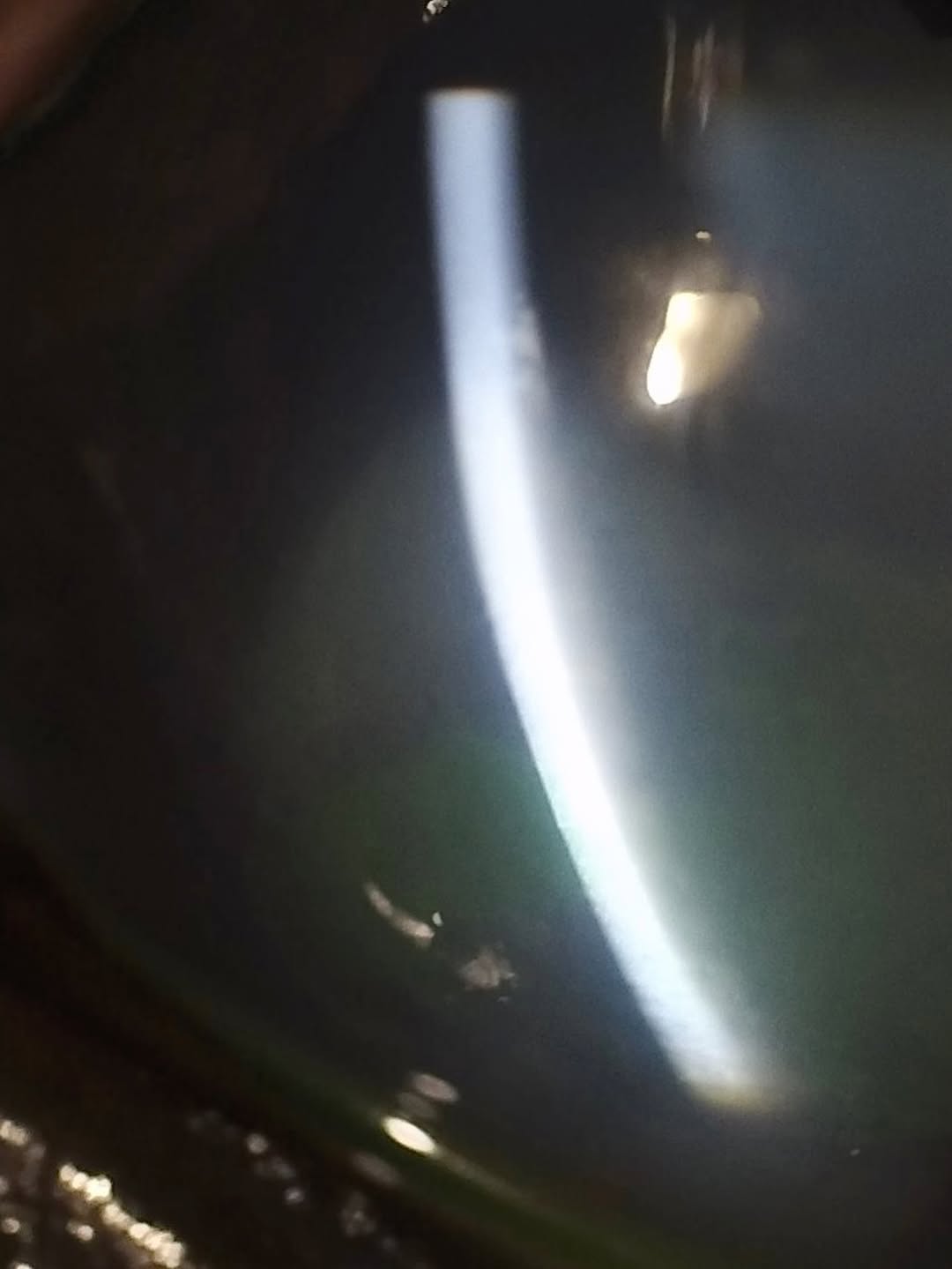
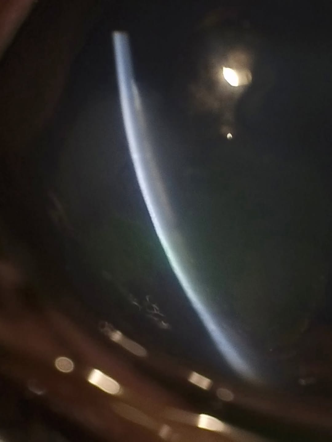
The first photo shows the whole cornea and the ulcer. The next two use the slit lamp imaging on the @kowaamericancorp SL-19 to show how thick the cornea gets in the affected area! Pretty cool to be able to show, and useful when doing exams with owners and primary care veterinarians. They can watch the whole thing on a tablet or phone screen while I am using the slit lamp!
 |  |  |
We are glad to share a vet case study captured by QuikVue eye imaging adaptor from Dr. Allison Fuchs.
This is a tough case! This is a horse with corneal ulcers that keep coming back over an area of corneal edema, which just means fluid in the cornea. They have been battling this for several years and we just started seeing him.
There are several possible causes for focal corneal edema in horses, including a form of immune mediated disease, traumatic damage to or degeneration of the inner layer of the cornea, or rare forms of uveitis. Once we get this ulcer healed we are going to see if we can keep this from happening so often!
The first photo shows the whole cornea and the ulcer. The next two use the slit lamp imaging on the @kowaamericancorp SL-19 to show how thick the cornea gets in the affected area! Pretty cool to be able to show, and useful when doing exams with owners and primary care veterinarians. They can watch the whole thing on a tablet or phone screen while I am using the slit lamp!
| | |