AnterVue: Turn Your Smartphone Into a Practical Anterior Segment Imaging Tool
For many optometrists, efficiency matters just as much as diagnostic quality. During a busy clinic day, having a fast and convenient way to capture anterior segment images can improve workflow, strengthen patient communication, and support better record keeping.
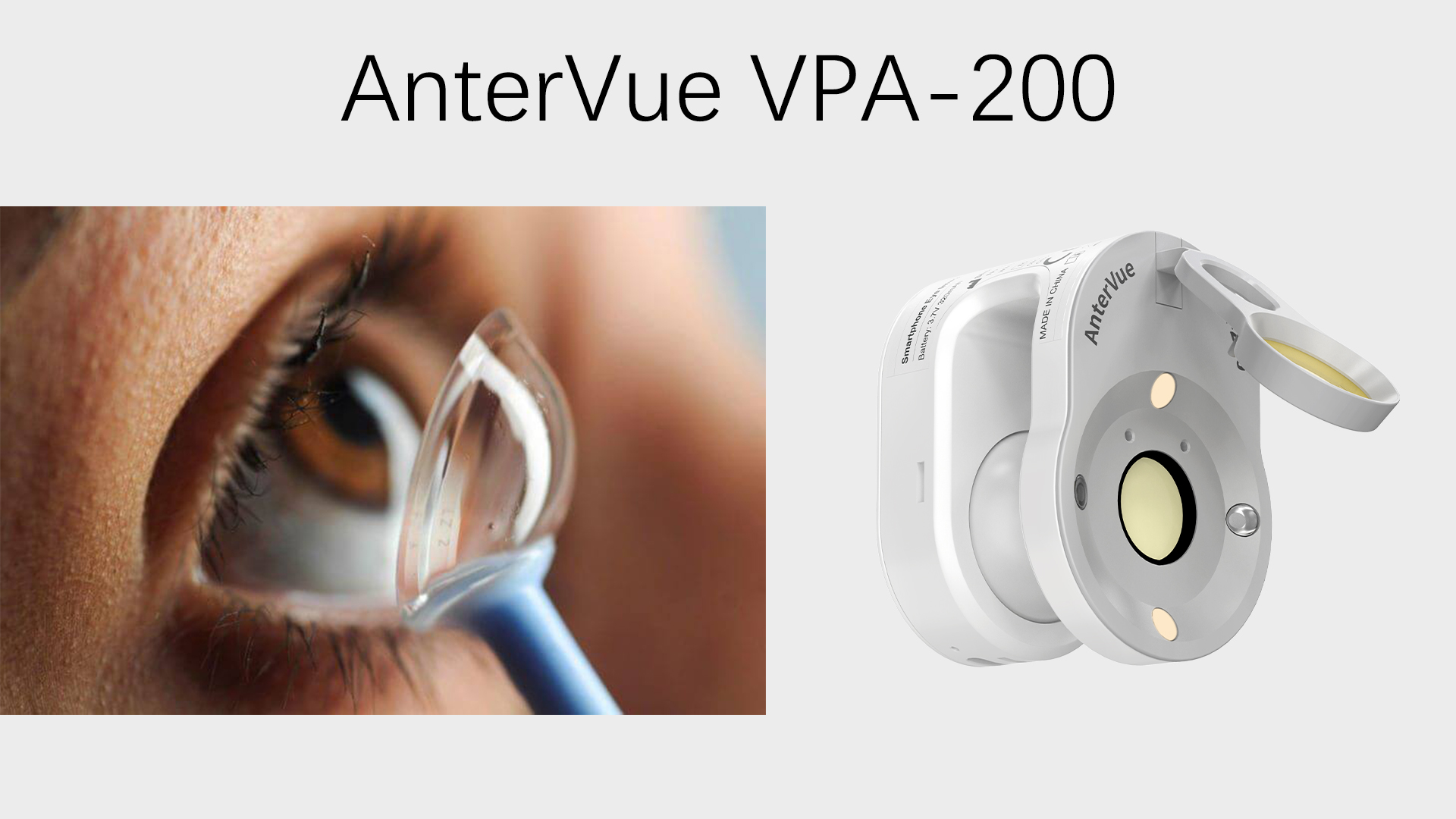
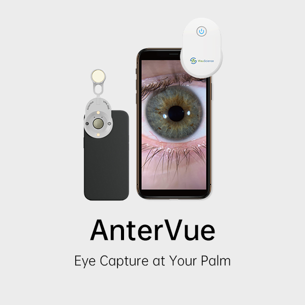
That is where AnterVue comes in. AnterVue (previously known as QuikVue) is a portable smartphone eye imaging adaptor designed to help eye care professionals capture clear anterior segment images using the smartphone they already carry every day.
High-Quality Imaging With Your Smartphone
AnterVue attaches to a smartphone or tablet and utilizes the device’s main camera to take detailed images of the eye’s anterior segment. This gives ODs a convenient option for documenting findings without relying on a dedicated digital slit lamp camera.
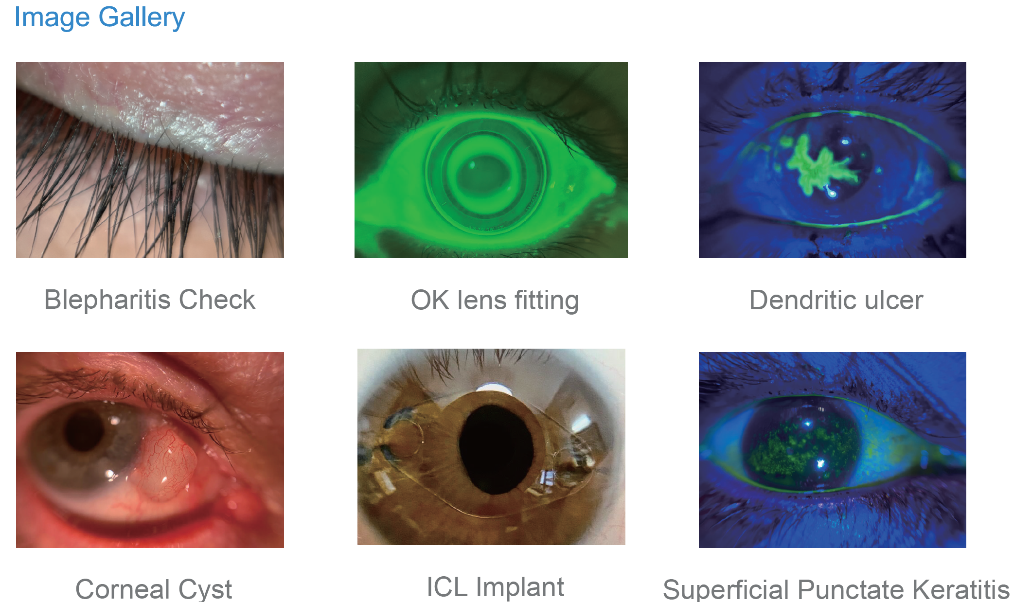
It is ideal for imaging:
- Corneal abnormalities
- Conjunctival findings
- Eyelid conditions
- Ocular surface changes
- Contact lens fitting observations
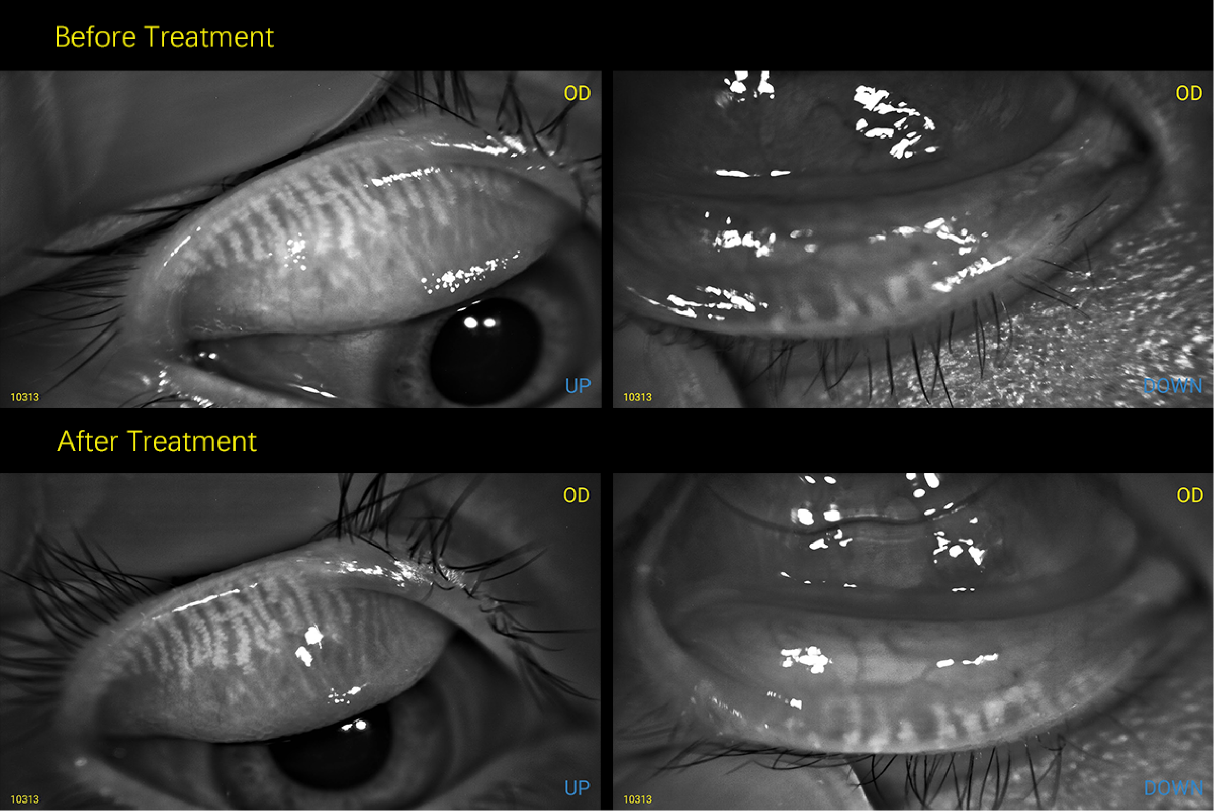
- Post-treatment follow-up comparisons
10X Magnification for Clear Clinical Detail
AnterVue features a 10X magnification lens, helping practitioners capture close-up images with the level of detail needed for everyday clinical communication and documentation.
Whether reviewing lid margins, corneal lesions, or surface staining, magnified imaging can make subtle findings easier to visualize and explain.
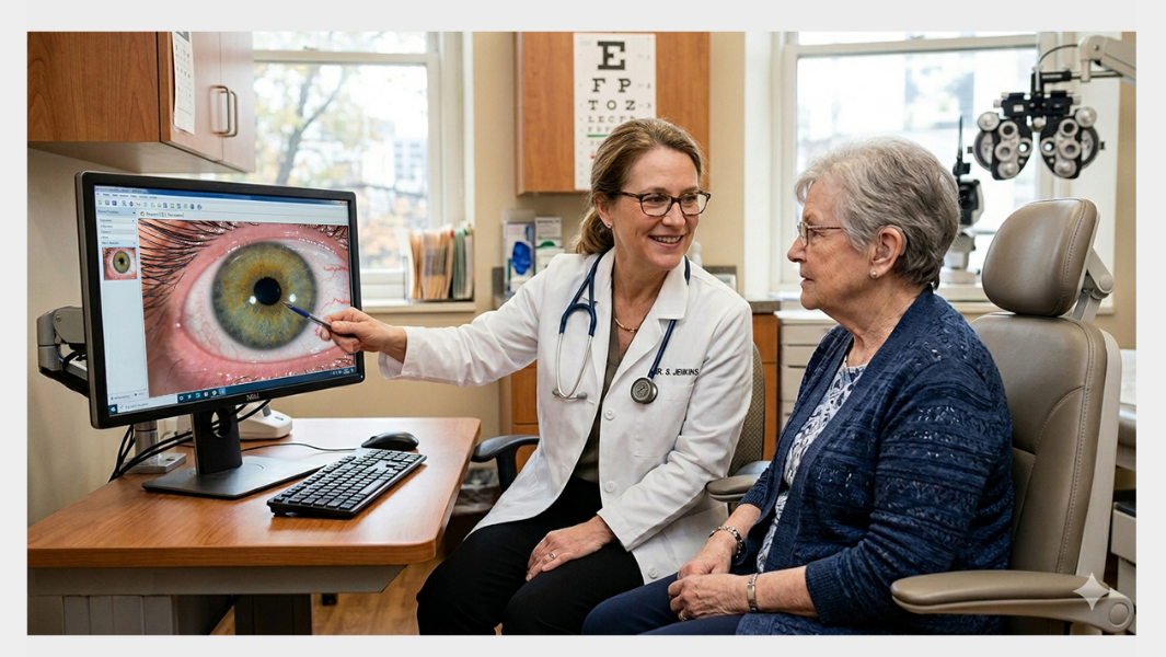
Improve Patient Education Instantly
Patients often understand their condition better when they can see it. Instead of only describing blepharitis, corneal staining, or redness, ODs can show real images captured during the exam.
Visual education can improve:
- Treatment acceptance
- Compliance with lid hygiene or dry eye therapy
- Follow-up engagement
- Trust in diagnosis and recommendations
A Smart Daily Tool for ODs
For optometrists who want a practical and affordable way to add imaging to routine care, AnterVue offers a strong balance of portability, convenience, and clinical usefulness.
Instead of waiting for access to a camera-equipped slit lamp, doctors can capture images immediately with the smartphone already in hand.
User Feedback
Hear how one clinic owner shares her experience using AnterVue in a small practice setting. She explains how this convenient smartphone imaging tool helps capture clear anterior segment photos, improve patient education, and streamline daily workflow.
How to Purchase
Interested doctors can visit www.dryeyestartup.com to purchase AnterVue and explore more practical solutions for dry eye diagnosis, patient education, and clinical workflow improvement. Discover tools designed to help grow and modernize your practice.
For many optometrists, efficiency matters just as much as diagnostic quality. During a busy clinic day, having a fast and convenient way to capture anterior segment images can improve workflow, strengthen patient communication, and support better record keeping.
That is where AnterVue comes in. AnterVue (previously known as QuikVue) is a portable smartphone eye imaging adaptor designed to help eye care professionals capture clear anterior segment images using the smartphone they already carry every day.
High-Quality Imaging With Your Smartphone
AnterVue attaches to a smartphone or tablet and utilizes the device’s main camera to take detailed images of the eye’s anterior segment. This gives ODs a convenient option for documenting findings without relying on a dedicated digital slit lamp camera.
It is ideal for imaging:
- Corneal abnormalities
- Conjunctival findings
- Eyelid conditions
- Ocular surface changes
- Contact lens fitting observations
- Post-treatment follow-up comparisons
10X Magnification for Clear Clinical Detail
AnterVue features a 10X magnification lens, helping practitioners capture close-up images with the level of detail needed for everyday clinical communication and documentation.
Whether reviewing lid margins, corneal lesions, or surface staining, magnified imaging can make subtle findings easier to visualize and explain.
Improve Patient Education Instantly
Patients often understand their condition better when they can see it. Instead of only describing blepharitis, corneal staining, or redness, ODs can show real images captured during the exam.
Visual education can improve:
- Treatment acceptance
- Compliance with lid hygiene or dry eye therapy
- Follow-up engagement
- Trust in diagnosis and recommendations
A Smart Daily Tool for ODs
For optometrists who want a practical and affordable way to add imaging to routine care, AnterVue offers a strong balance of portability, convenience, and clinical usefulness.
Instead of waiting for access to a camera-equipped slit lamp, doctors can capture images immediately with the smartphone already in hand.
User Feedback
Hear how one clinic owner shares her experience using AnterVue in a small practice setting. She explains how this convenient smartphone imaging tool helps capture clear anterior segment photos, improve patient education, and streamline daily workflow.
How to Purchase
Interested doctors can visit www.dryeyestartup.com to purchase AnterVue and explore more practical solutions for dry eye diagnosis, patient education, and clinical workflow improvement. Discover tools designed to help grow and modernize your practice.