How to Interpret Meibography
When interpreting a meibography, it is important to grade the quality and quantity of the glands by analyzing three key factors: atrophy, tortuosity and segmentation, each of which can be graded from 0 to 4.
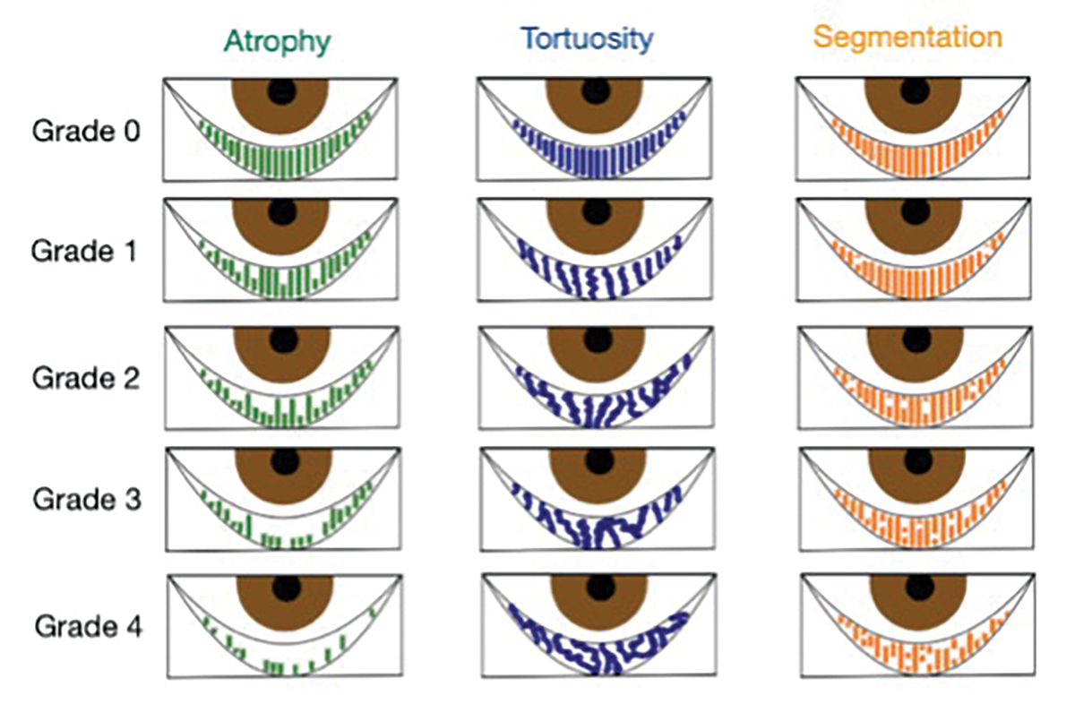 |
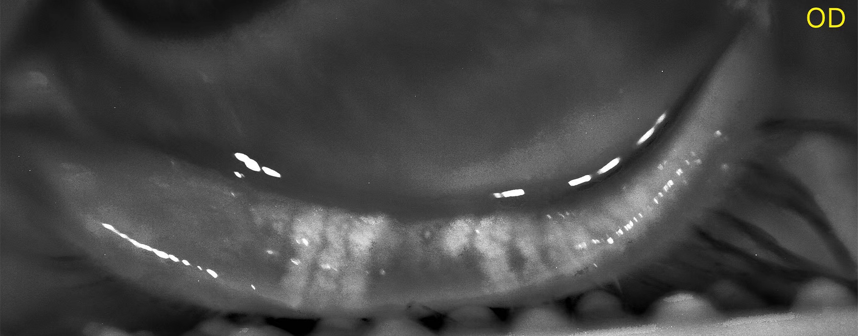
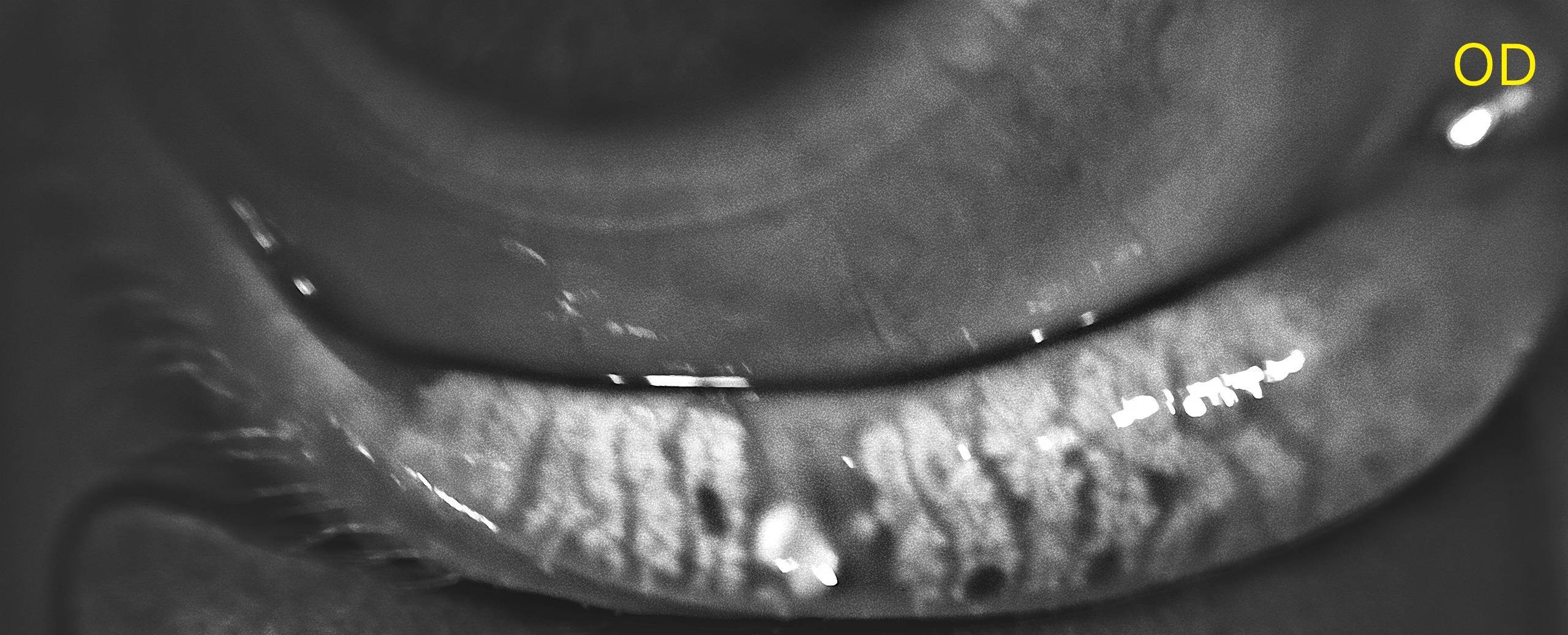
Atrophy, described by the Pult 5-grade scale, measures the loss of meibomian glands, ranging from partial to complete loss from the orifice to the fornix.
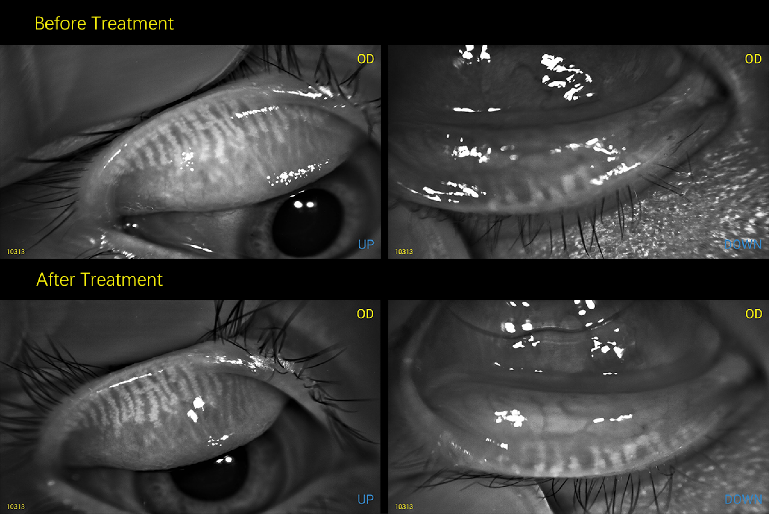
Image examples captured by MeiboVue:
 |
|
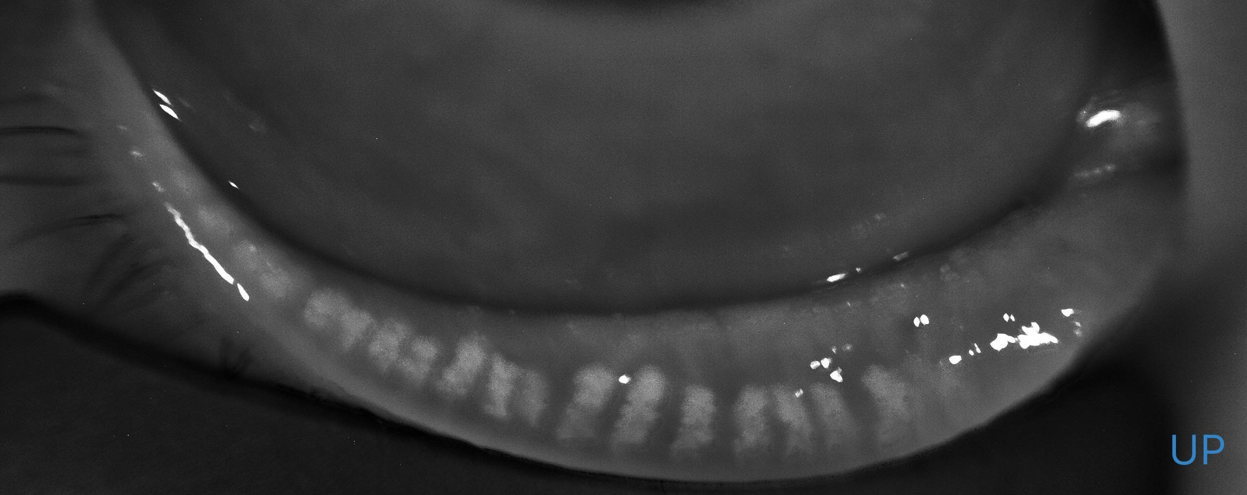
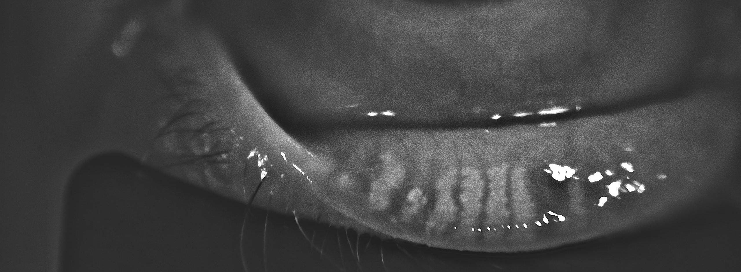
Tortuosity, assessed using the Halleran grading scale, is present if a gland bends at a 45º angle from the midline or if there are multiple bends of any degree within a gland.
Image examples captured by MeiboVue:
 |
|
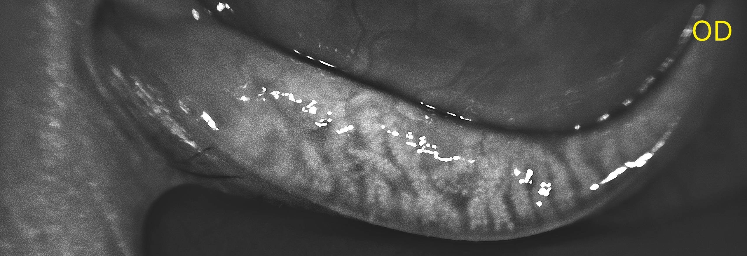
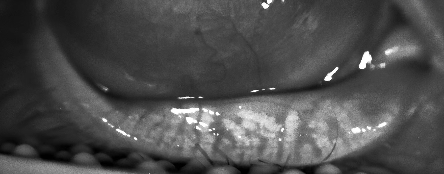
Segmentation, evaluated using a segmentation grading scale, describes the broken appearance of the glands, often indicated by a division within a gland marked by a black line.
Image examples captured by MeiboVue:
 |
|
These grading systems provide a quick and standardized method for documenting the quality of meibomian glands as shown on the meibography.
Original source:
https://www.revieweducationgroup.com/ce/a-stepwise-approach-to-diagnosing-dry-eye-disease
When interpreting a meibography, it is important to grade the quality and quantity of the glands by analyzing three key factors: atrophy, tortuosity and segmentation, each of which can be graded from 0 to 4.
|
Atrophy, described by the Pult 5-grade scale, measures the loss of meibomian glands, ranging from partial to complete loss from the orifice to the fornix.
Image examples captured by MeiboVue:
|
|
Tortuosity, assessed using the Halleran grading scale, is present if a gland bends at a 45º angle from the midline or if there are multiple bends of any degree within a gland.
Image examples captured by MeiboVue:
|
|
Segmentation, evaluated using a segmentation grading scale, describes the broken appearance of the glands, often indicated by a division within a gland marked by a black line.
Image examples captured by MeiboVue:
|
|
These grading systems provide a quick and standardized method for documenting the quality of meibomian glands as shown on the meibography.
Original source:
https://www.revieweducationgroup.com/ce/a-stepwise-approach-to-diagnosing-dry-eye-disease