QuikVue Vet Case Share - abscess in the cornea
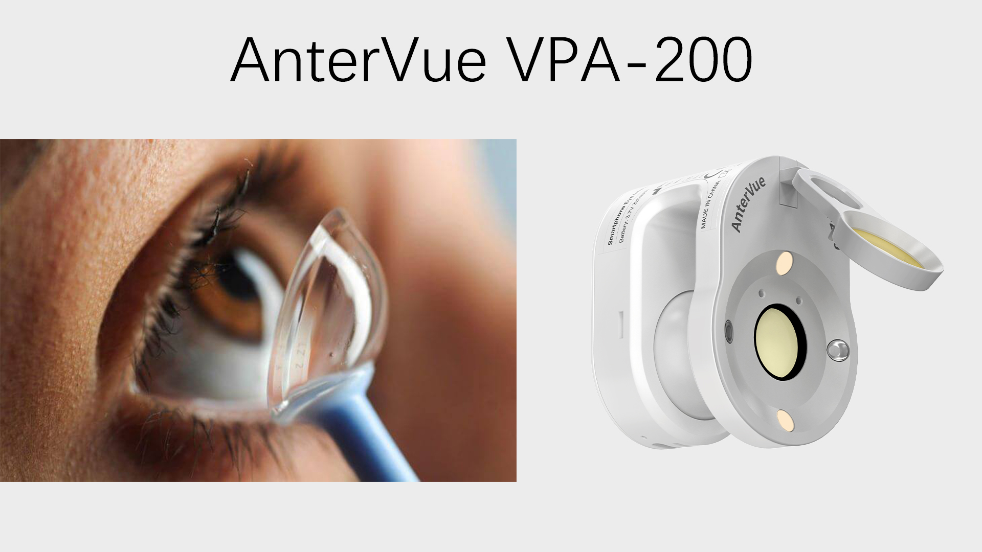
We are glad to share a vet case study captured by QuikVue eye imaging adaptor from Dr. Allison Fuchs.
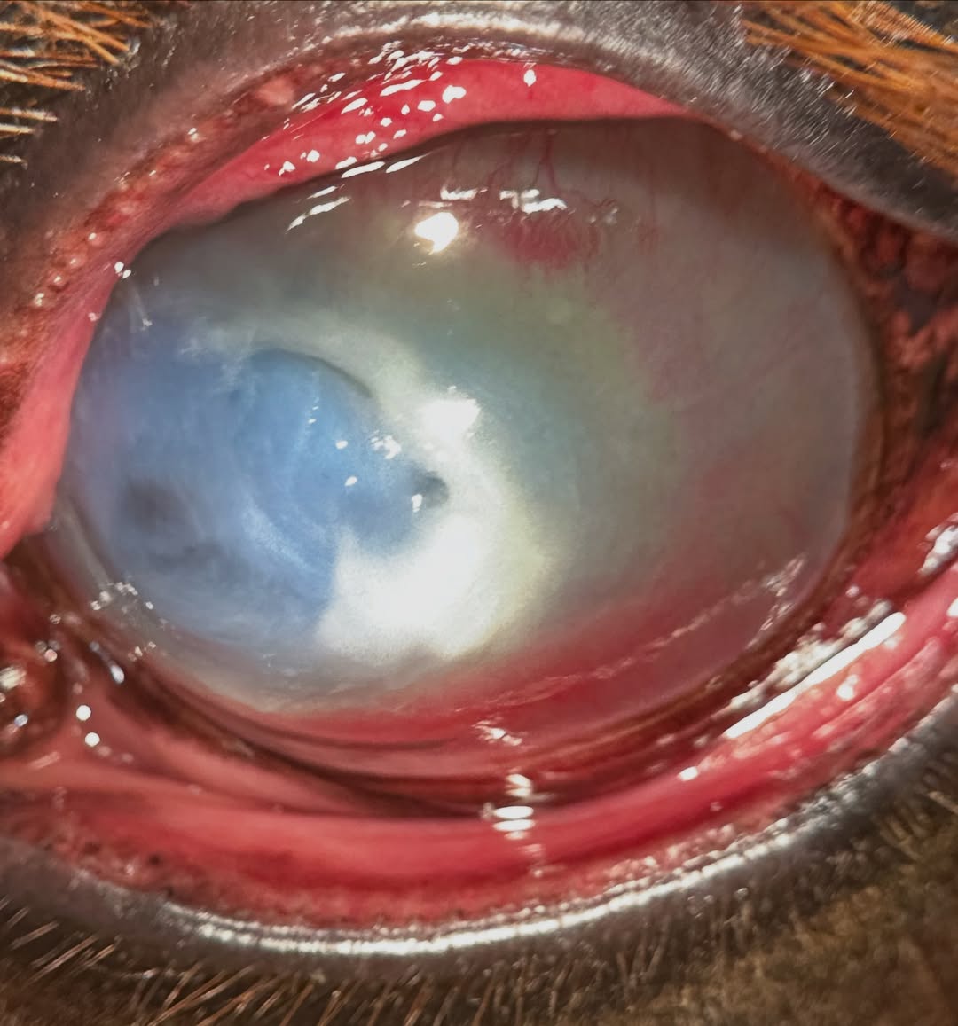
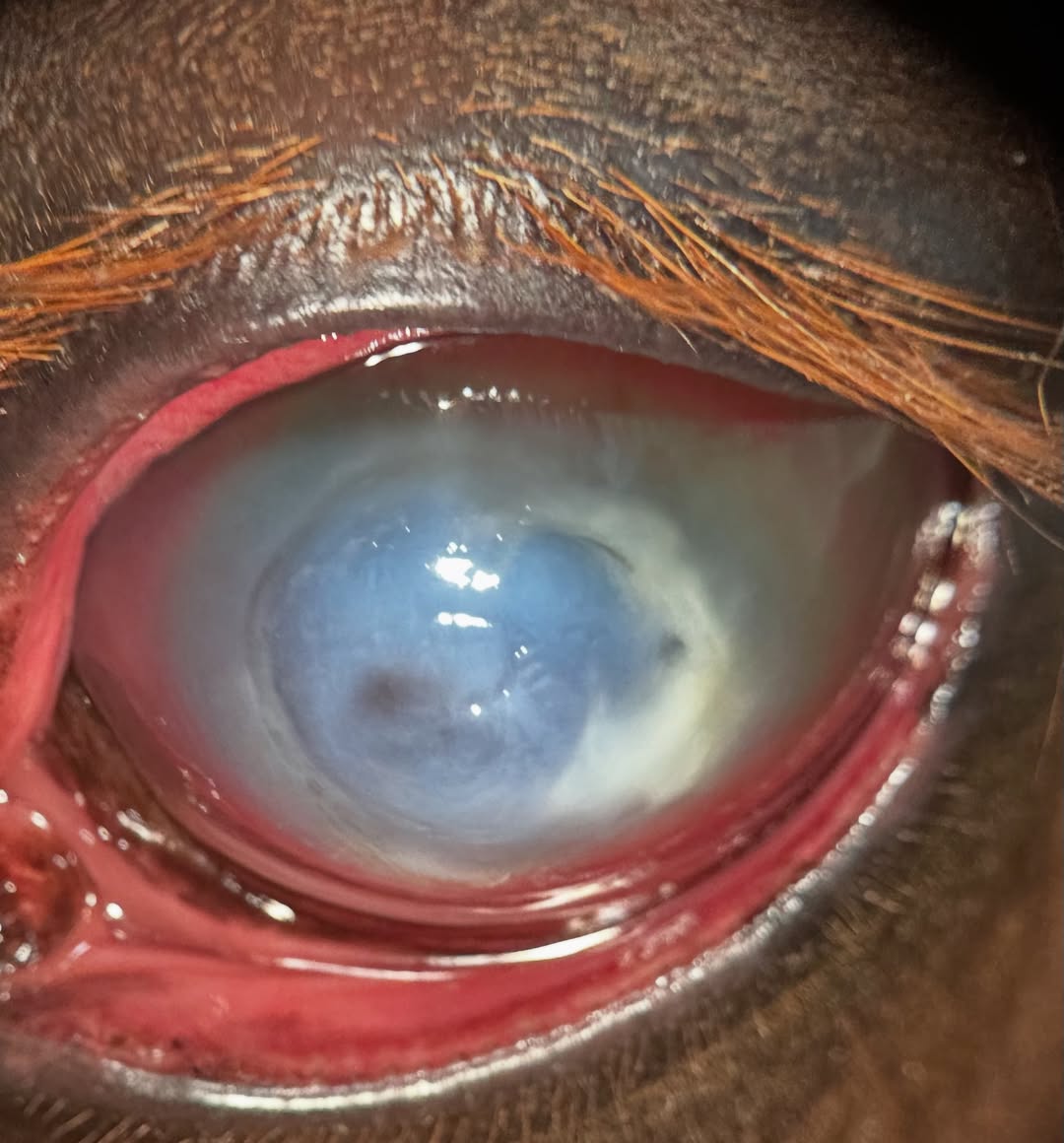
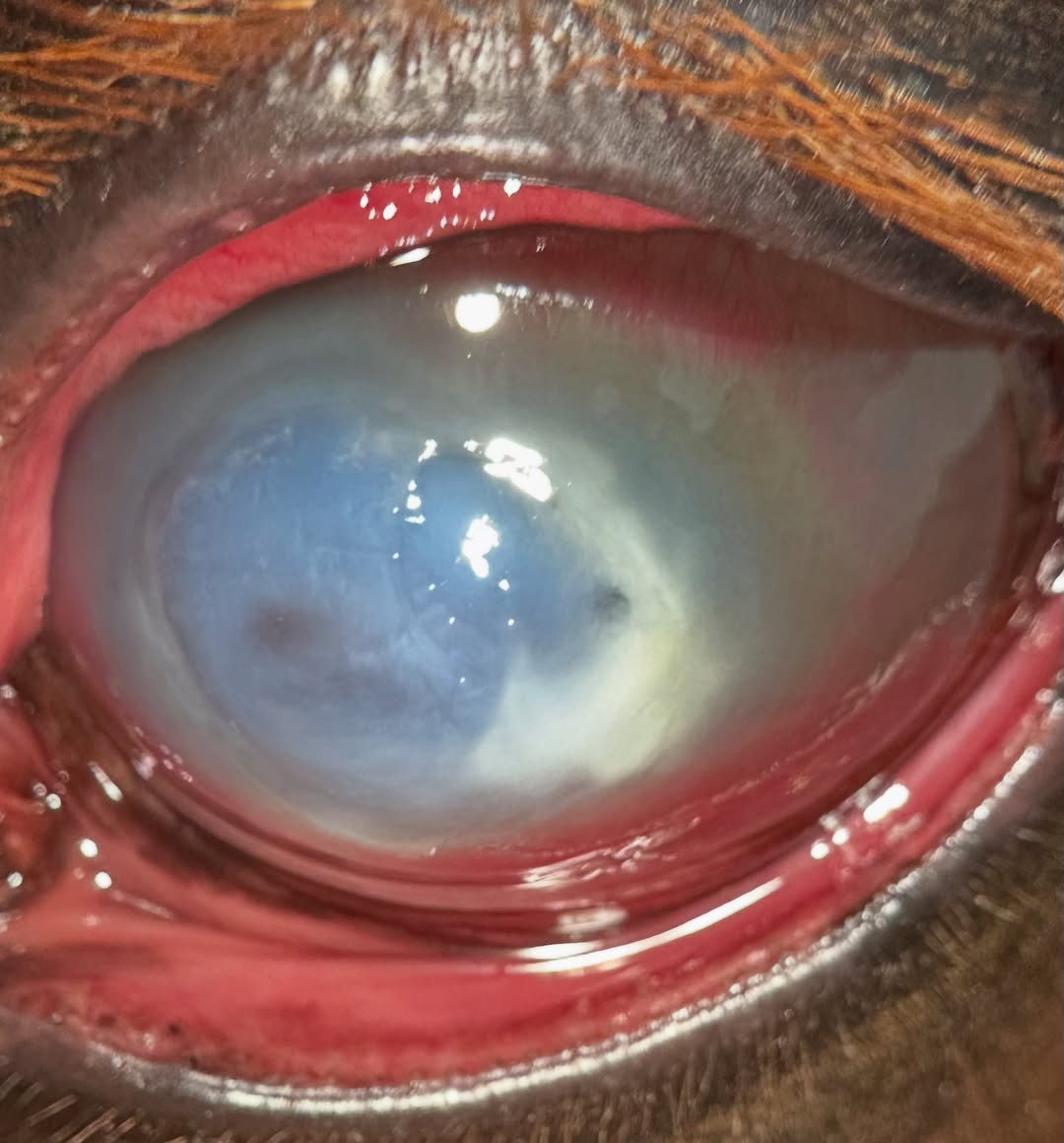
We have just been swimming in fungal keratitis lately! Today I saw another horse with an abscess in the cornea that has been unresponsive to treatment. This horse had an unknown injury 2-3 weeks ago, and was being treated with topical eye ointments.
Normally I try to get samples for cytology, but the epithelium has completely sealed over here, making the infection inaccessible without opening the corneal tissue again. However, most of the time we know this is fungal in origin due to the part of the world we live in and the prevalence in horses. We have to use medications that penetrate the intact corneal epithelium if we aren’t going to perform surgery, which wasn’t an option in this case. We placed a subpalpebral lavage tube and have started the horse on aggressive topical medication. This can be a long course of treatment, lasting weeks, and the prognosis is still guarded until we see how they respond.
 |  |  |
We are glad to share a vet case study captured by QuikVue eye imaging adaptor from Dr. Allison Fuchs.
We have just been swimming in fungal keratitis lately! Today I saw another horse with an abscess in the cornea that has been unresponsive to treatment. This horse had an unknown injury 2-3 weeks ago, and was being treated with topical eye ointments.
Normally I try to get samples for cytology, but the epithelium has completely sealed over here, making the infection inaccessible without opening the corneal tissue again. However, most of the time we know this is fungal in origin due to the part of the world we live in and the prevalence in horses. We have to use medications that penetrate the intact corneal epithelium if we aren’t going to perform surgery, which wasn’t an option in this case. We placed a subpalpebral lavage tube and have started the horse on aggressive topical medication. This can be a long course of treatment, lasting weeks, and the prognosis is still guarded until we see how they respond.
| | |