




QuikVue Vet Case Share - retinal detachment
We are glad to share a vet case study captured by QuikVue eye imaging adaptor from Dr. Allison Fuchs.
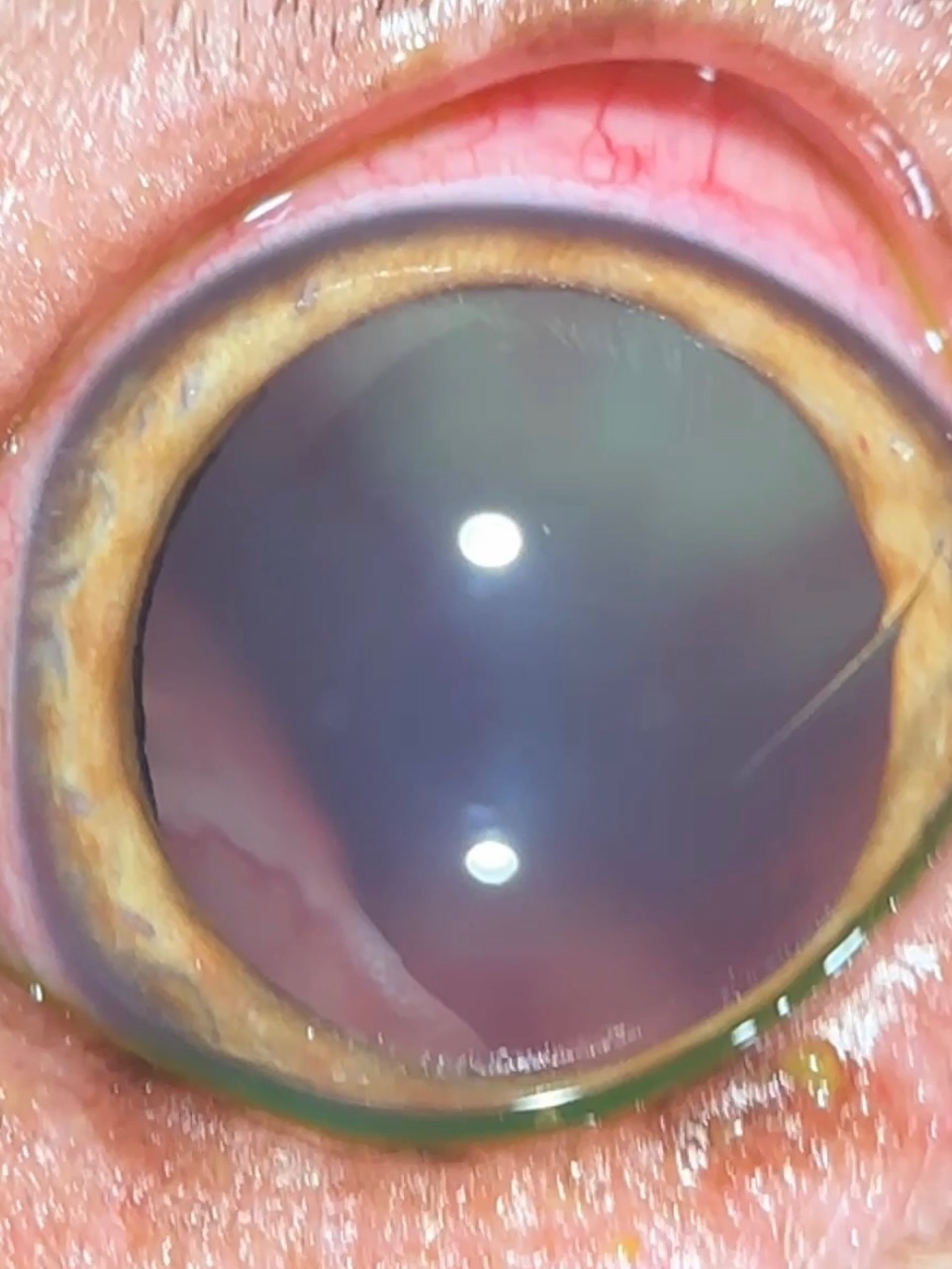
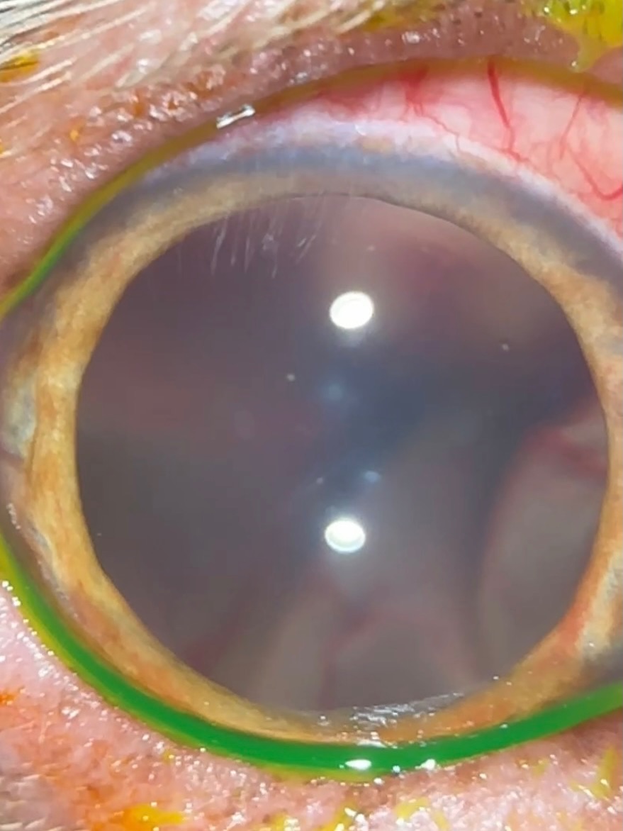
A reminder that if you can see retinal blood vessels through the pupil like this without a fundoscopy lens, you have a retinal detachment!
This dog presented to the ER for acute vision loss, and was found to have dilated pupils. On ophthalmic exam, we found uveitis (inflammation in the anterior chamber) and chorioretinitis (inflammation in the retina) with bullous retinal detachments.
This can be due to systemic infections (fungal, tick-borne, etc), neoplasia (cancer), or immune mediated causes. A systemic workup is recommended to look for underlying diseases that would need to be treated, and if those causes are ruled out, these can be treated as immune-mediated with immunosuppressants. With appropriate diagnostics and therapy, many of these dogs can have retinal reattachment with resolution of disease - meaning they regain vision!
 |  |
We are glad to share a vet case study captured by QuikVue eye imaging adaptor from Dr. Allison Fuchs.
A reminder that if you can see retinal blood vessels through the pupil like this without a fundoscopy lens, you have a retinal detachment!
This dog presented to the ER for acute vision loss, and was found to have dilated pupils. On ophthalmic exam, we found uveitis (inflammation in the anterior chamber) and chorioretinitis (inflammation in the retina) with bullous retinal detachments.
This can be due to systemic infections (fungal, tick-borne, etc), neoplasia (cancer), or immune mediated causes. A systemic workup is recommended to look for underlying diseases that would need to be treated, and if those causes are ruled out, these can be treated as immune-mediated with immunosuppressants. With appropriate diagnostics and therapy, many of these dogs can have retinal reattachment with resolution of disease - meaning they regain vision!
| |