QuikVue Vet Case Share-a deep ulcer
We are glad to share a vet case study captured by QuikVue eye imaging adaptor from Dr. Allison Fuchs.
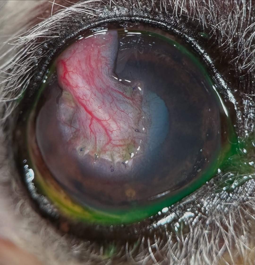
Progress update! I have showed you this dog before and he was just in for his (hopefully) final recheck with me. This sweet pup initially came for a deep ulcer and the owners wanted to avoid surgery due to his age. Since it hadn’t been on any treatment and didn’t appear infected, we started therapy, but just 48 hours later it tanked into a very gnarly infected descemetocele. We performed a keratectomy to remove as much of the infected cornea as possible, and placed a conjunctival graft for tectonic support and to bring in blood vessels. The graft can remain in place forever! Here you can see his progress from initial exam, 48 hours later when we decided to go to surgery, 1 week recheck post-graft, 3 weeks post-surgery, and finally 6 weeks post-operative.
Little man is visual, comfortable, and has been back to his normal routine for a while. These guys are usually in a cone for at least 2 weeks (sometimes more), but stabilizing the cornea and saving the eye and vision is worth it for most of our owners! Due to his age and breed, we have elected not to trim the graft, but in another few months it will be even more scarred and less noticeable! Since they don’t have to read emails or drive, the visual impact is much less significant than it would be for a person. Pretty neat what we can achieve!
 | |
We are glad to share a vet case study captured by QuikVue eye imaging adaptor from Dr. Allison Fuchs.
Progress update! I have showed you this dog before and he was just in for his (hopefully) final recheck with me. This sweet pup initially came for a deep ulcer and the owners wanted to avoid surgery due to his age. Since it hadn’t been on any treatment and didn’t appear infected, we started therapy, but just 48 hours later it tanked into a very gnarly infected descemetocele. We performed a keratectomy to remove as much of the infected cornea as possible, and placed a conjunctival graft for tectonic support and to bring in blood vessels. The graft can remain in place forever! Here you can see his progress from initial exam, 48 hours later when we decided to go to surgery, 1 week recheck post-graft, 3 weeks post-surgery, and finally 6 weeks post-operative.
Little man is visual, comfortable, and has been back to his normal routine for a while. These guys are usually in a cone for at least 2 weeks (sometimes more), but stabilizing the cornea and saving the eye and vision is worth it for most of our owners! Due to his age and breed, we have elected not to trim the graft, but in another few months it will be even more scarred and less noticeable! Since they don’t have to read emails or drive, the visual impact is much less significant than it would be for a person. Pretty neat what we can achieve!
| |