




QuikVue Vet Case Share-indolent corneal ulcer
We are glad to share a vet case study captured by QuikVue eye imaging adaptor from Dr. Allison Fuchs.
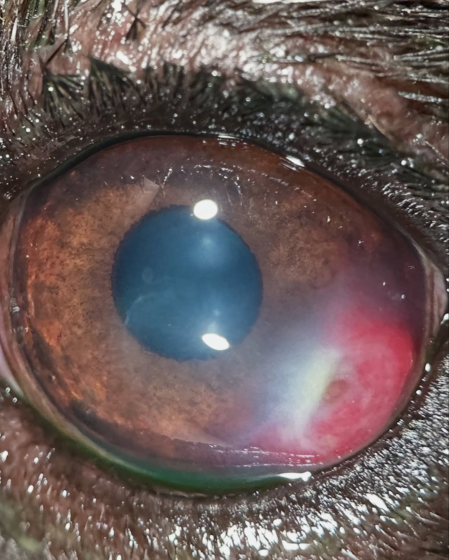
This sweet Frenchie had recently gotten over an indolent corneal ulcer in the OTHER eye when her mom rushed her in for an urgent appointment due to squinting the left eye. Somehow, Winni had managed to develop a very severe corneal infection! Over 48 hours it progressed to a deep ulcer (second photo) and the infection wasn’t responding to topical and systemic antibiotics. We took Winni to surgery to perform a keratectomy, which means removing the affected area of cornea.
This allowed me to remove all the infected tissue, as well as get better samples for cytology and culture. The resulting corneal defect was grafted with BioSis, which is a bioscaffold material we can use for physical support of the cornea. Generally I use it with a conjunctival graft, but since the area had lots of blood vessels I did a “naked” biosis graft.
The third photo shows Winni 1 week later, with even more vessels but no infection! 2 more weeks and the 4th photo, and you can see everything is getting less inflamed as the graft absorbs and the vessels regress. Her final recheck was last week and she looks amazing! Both Winni’s mom and I are so pleased with her cosmetic and visual outcome. Way to go Winni!
 |  |
We are glad to share a vet case study captured by QuikVue eye imaging adaptor from Dr. Allison Fuchs.
This sweet Frenchie had recently gotten over an indolent corneal ulcer in the OTHER eye when her mom rushed her in for an urgent appointment due to squinting the left eye. Somehow, Winni had managed to develop a very severe corneal infection! Over 48 hours it progressed to a deep ulcer (second photo) and the infection wasn’t responding to topical and systemic antibiotics. We took Winni to surgery to perform a keratectomy, which means removing the affected area of cornea.
This allowed me to remove all the infected tissue, as well as get better samples for cytology and culture. The resulting corneal defect was grafted with BioSis, which is a bioscaffold material we can use for physical support of the cornea. Generally I use it with a conjunctival graft, but since the area had lots of blood vessels I did a “naked” biosis graft.
The third photo shows Winni 1 week later, with even more vessels but no infection! 2 more weeks and the 4th photo, and you can see everything is getting less inflamed as the graft absorbs and the vessels regress. Her final recheck was last week and she looks amazing! Both Winni’s mom and I are so pleased with her cosmetic and visual outcome. Way to go Winni!
| |