QuikVue Vet Case Study—Cataracts and Dry Eye in a Diabetic Dog
We are glad to share a vet case study captured by QuikVue eye imaging adaptor from Dr. Allison Fuchs.
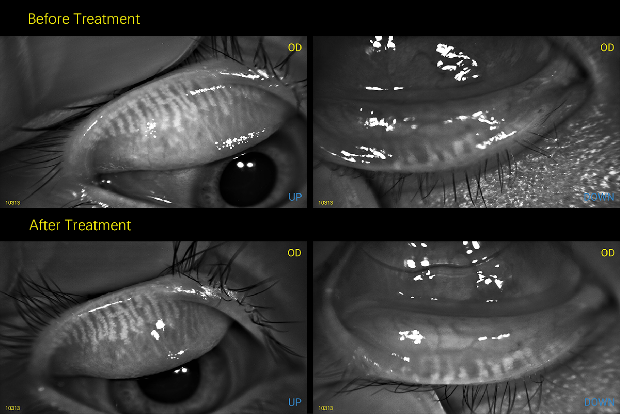
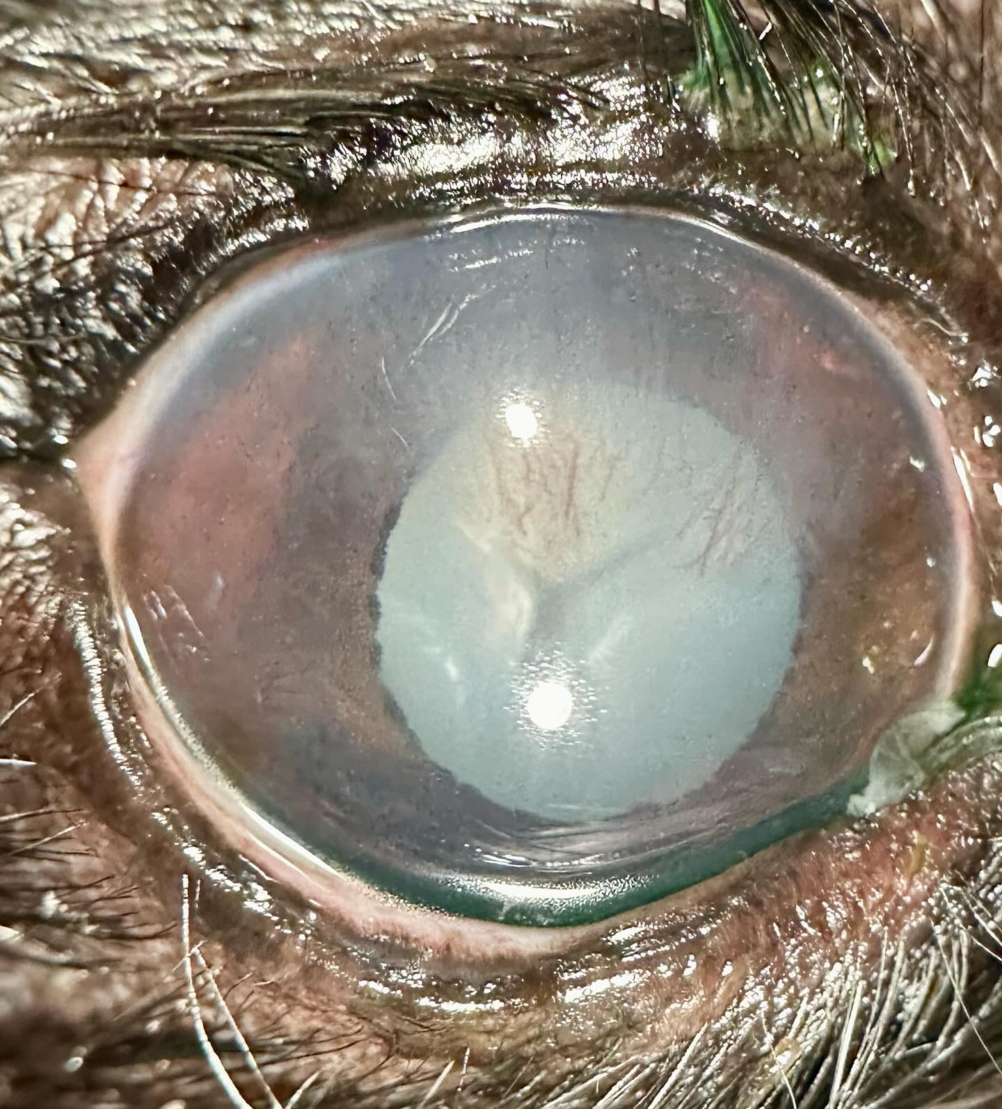
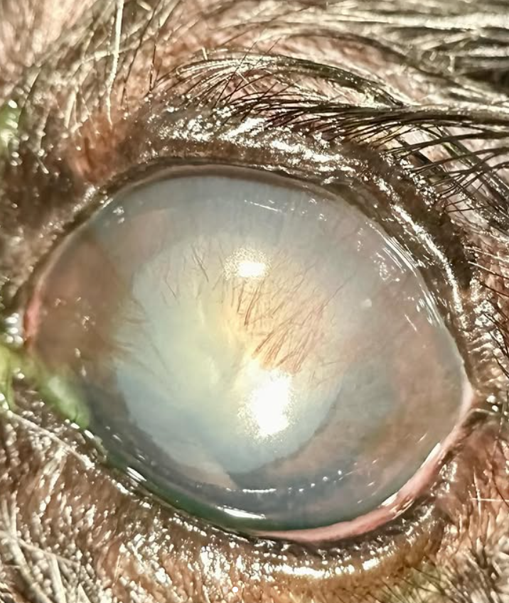
Take a look at these images and tell me what you think is going on with this dog before reading onwards. How many issues can you spot? What if I told you there was a big, invisible systemic issue too? This dog came to see me for vision loss and ocular discharge. The vision loss was acute, but the discharge had been going on for at least a few weeks. On exam, this dog had very low tear production (dry eye), which is the cause for the discharge and the corneal blood vessels you can see growing in from the top of the eye. We also diagnosed complete cataracts, which appeared very swollen (aka intumescent). At this point I was very suspicious of an underlying issue. More questioning revealed that the dog had been acting more lethargic, drinking and urinating more, and had lost weight over the past month. We checked bloodwork because I was concerned we were missing the big picture, and the dog’s blood glucose was nearly 500! This little dog had developed diabetes, but because the signs were fairly vague and insidious, hadn’t been diagnosed yet. Diabetes can be life threatening if untreated, so we immediately got them set up with insulin and a follow up plan for the dog’s ongoing care. About 85% of dogs with diabetes develop cataracts within the first year of being diabetic - while it’s uncommon for the ophthalmologist to find the diabetes, this is not the first time! Dry eye is also more common in diabetic dogs. I also started them on eye medication to help decrease inflammation and improve tear production. We are going to revisit and discuss options for cataract surgery once they have the diabetes a little better controlled.
 |  |
We are glad to share a vet case study captured by QuikVue eye imaging adaptor from Dr. Allison Fuchs.
Take a look at these images and tell me what you think is going on with this dog before reading onwards. How many issues can you spot? What if I told you there was a big, invisible systemic issue too? This dog came to see me for vision loss and ocular discharge. The vision loss was acute, but the discharge had been going on for at least a few weeks. On exam, this dog had very low tear production (dry eye), which is the cause for the discharge and the corneal blood vessels you can see growing in from the top of the eye. We also diagnosed complete cataracts, which appeared very swollen (aka intumescent). At this point I was very suspicious of an underlying issue. More questioning revealed that the dog had been acting more lethargic, drinking and urinating more, and had lost weight over the past month. We checked bloodwork because I was concerned we were missing the big picture, and the dog’s blood glucose was nearly 500! This little dog had developed diabetes, but because the signs were fairly vague and insidious, hadn’t been diagnosed yet. Diabetes can be life threatening if untreated, so we immediately got them set up with insulin and a follow up plan for the dog’s ongoing care. About 85% of dogs with diabetes develop cataracts within the first year of being diabetic - while it’s uncommon for the ophthalmologist to find the diabetes, this is not the first time! Dry eye is also more common in diabetic dogs. I also started them on eye medication to help decrease inflammation and improve tear production. We are going to revisit and discuss options for cataract surgery once they have the diabetes a little better controlled.
| |